One of the biggest causes of death on the globe is heart disease. Timely diagnosis and appropriate treatment can significantly improve the patient’s prognosis. Myocardial perfusion imaging (MPI) is a powerful diagnostic tool used to evaluate blood flow in the heart muscle and detect problems such as coronary artery disease. In this blog, we’ll dive deep into the world of MPI testing, exploring what it is, why it’s done, how it works, and what to expect during the process.
1. What is the MPI Test?
Myocardial perfusion imaging (MPI) is a non-invasive imaging technique used to assess blood flow in the heart muscle. It provides essential information about the heart’s function, particularly its ability to receive adequate blood supply. The test is particularly useful in diagnosing and determining the severity of coronary artery disease (CAD).
2. Why is the MPI test done?
MPI tests are usually conducted for the following reasons:
Diagnosing Coronary Artery Disease (CAD): CAD occurs when the coronary arteries, which supply blood to the heart, become narrowed or blocked. MPI can help identify areas of reduced blood flow to the heart muscle, indicating CAD.
Assessing heart function: MPI can assess how well your heart is pumping blood and identify any areas of poor muscle function, which may be due to a lack of blood flow.
Evaluation of the effect of heart treatment: It can also be used to evaluate the effectiveness of previous treatments such as angioplasty or coronary artery bypass surgery. Determining heart attack risk: For people at risk for heart disease, the MPI can provide valuable information about future heart attack risk.
3. How does the MPI test work?
Two methods are generally used in MPI: stress imaging and rest imaging. Here’s how the test works:
Stress imaging: In this phase, you will be exposed to stress, either through exercise on a treadmill or by administering drugs that mimic the effects of exercise. Stress increases your heart rate and blood flow, allowing doctors to assess how well your heart responds to stress. A radioactive chemical known as a radiotracer is put into your circulation in a very tiny quantity. The blood carries this tracer to the heart muscle. Healthy heart muscle takes up the tracer equally, whereas areas with reduced blood flow take up less.
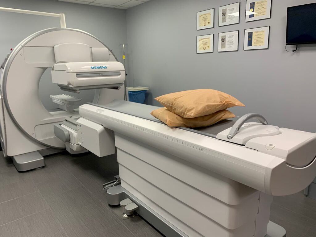
Imaging: Special cameras called gamma cameras are used to take pictures of the heart, capturing the distribution of radiotracers. These images help identify areas of decreased blood flow and muscle function.
Rest imaging: After the stressor, you will be asked to rest for some time. Another set of images is taken, which allows doctors to compare your heart’s blood flow and function at rest with its function during stress.
4. What to expect during the process:
The MPI test is usually performed in a hospital or specialized imaging center. Before the test, you might need to fast or abstain from caffeine. You will be connected to an ECG machine to monitor your heart’s electrical activity throughout the test. The radiotracer is injected intravenously. If you are undergoing a stress test, you may be asked to exercise on a treadmill or ride a stationary bike. The entire procedure may take several hours.
5. Interpreting MPI Results:
Images obtained from MPI are carefully analyzed by radiologists or cardiologists. The results will provide important information, including Perfusion defect: Areas of reduced blood flow to the heart muscle will be identified. These are often referred to as “perfusion defects” and may indicate the presence of coronary artery disease or other heart conditions. Ejection fraction: The ejection fraction of your heart, which gauges how well it pumps blood, may also be determined by MPI. A reduced ejection fraction can be a sign of heart failure.
Comparative analysis: Images obtained during the stress and rest phases will be compared to assess any differences in blood flow or muscle function. This comparison helps to diagnose heart conditions and determine their severity.
6. Follow-up and treatment
Depending on the results of the MPI test, your healthcare provider will discuss the appropriate next steps. Possible outcomes and recommendations may include:
General results: A normal MPI test indicates that your heart is getting adequate blood flow and there are no significant blockages or areas of concern. In this case, your doctor may recommend ongoing heart-healthy lifestyle choices and regular check-ups. Results that are abnormal: If the MPI test reveals variations in blood flow or muscle performance, more testing and therapy may be required. Treatment options may include medications, lifestyle changes, angioplasty, stent placement, or coronary artery bypass surgery.
7. Risks and Safety Concerns:
MPI tests are generally safe, but as with any medical procedure, there are some risks and safety considerations to keep in mind:
Radiation exposure: Radiotracers used in MPI emit small amounts of radiation. Although the levels are generally considered safe, it is essential to inform your healthcare provider if you are pregnant or breastfeeding, as radiation exposure could potentially harm a developing fetus or baby.
Allergic reactions: Although rare, some people may have allergic reactions to radiotracer injections. Tell your healthcare team if you have a history of allergies or have experienced adverse drug reactions in the past.
Exercise stress test risks: If you undergo a stress test by exercising on a treadmill or stationary bike, there is a risk of cardiovascular complications such as arrhythmia or chest pain. However, healthcare professionals monitor your condition closely during the exam.
8. Future Directions:
The field of cardiac imaging is constantly evolving. Some possible future developments in the MPI test may include:
Artificial Intelligence (AI): AI-powered image analysis can increase the accuracy and efficiency of MPI interpretation. Machine learning algorithms can help detect subtle abnormalities in perfusion patterns and predict risk. Personalized medicine: MPI can be more personalized with treatment plans tailored to an individual’s specific perfusion and cardiac function characteristics. Non-radioactive tracers: Research is underway to develop non-radioactive tracers for MPI testing, further reducing radiation exposure